Posterior Shoulder Tendon Anatomy : What Causes Shoulder Pain | Shoulder Doctor | South ... - Classically associated with seizures and lightning strikes.
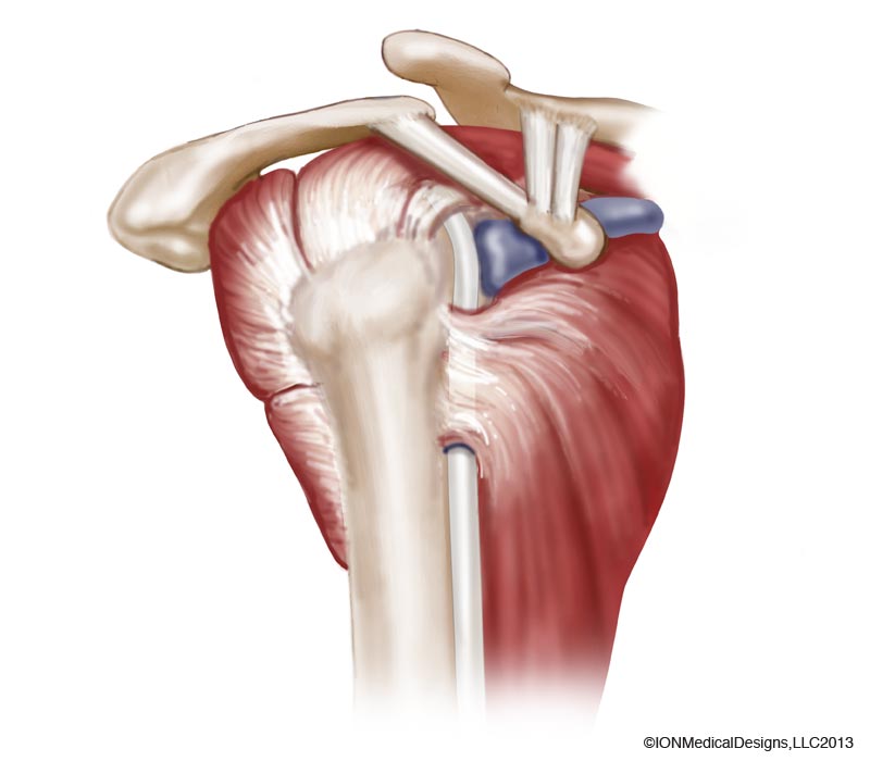
Posterior Shoulder Tendon Anatomy : What Causes Shoulder Pain | Shoulder Doctor | South ... - Classically associated with seizures and lightning strikes.. Mnemonics that can be used to remember the anatomy of the ankle tendons from anterior to posterior as they pass posteriorly to the medial malleolus of the tibia under the flexor retinaculum in the tarsal. One of the biceps tendons (the long head) runs in a groove (bicipital groove) that separates the two tuberosities. Anterior graphic of the shoulder. Posterior band of the ighl. The supraspinatus tendon and subacromial bursa).
Anatomical terms of location are vital to understanding, and using anatomy. Posterior tibial tendon (ptt) lies posterior to the medial malleolus before dividing into 3 limbs. Being an undergraduate student excites me and inspires me to lean. Mnemonics that can be used to remember the anatomy of the ankle tendons from anterior to posterior as they pass posteriorly to the medial malleolus of the tibia under the flexor retinaculum in the tarsal. In the shoulder it's commonly.
Anatomy of the Shoulder Archives - Joint Preservation Center from josephbermanmd.com Posterior tibial tendon dysfunction is a common problem of the foot and ankle. Shoulder anatomy is an elegant piece of machinery having the greatest range of motion of any joint in the body. Being an undergraduate student excites me and inspires me to lean. Posterior shoulder instability, accelerated osteoarthritis and pos the shoulder joint is functionally and structurally complex and is composed of bone, hyaline cartilage, labrum, ligaments objective: Aphrodite, athletic trainer, saint francis memorial hospital, demonstrates the anatomy of the posterior tibial tendon often injured for dr rich blake's blog. Shoulder is composed of 4 joints, namely glenohumeral or shoulder joint, acromioclavicular joint. Posterior graphic of the shoulder. Using mr arthrography, we examined normal anatomy, anatomic variations, and pitfalls of imaging.
Learn vocabulary, terms and more with flashcards, games and other study tools.
Classically associated with seizures and lightning strikes. Acute tears may occur when the arm is violently pushed into. Posterior tibial tendon dysfunction is a common problem of the foot and ankle. .posterior shoulder bone anatomy human shoulder joint anatomy frozen shoulder anatomy right shoulder muscle anatomy shoulder arm muscles anatomy shoulder anatomy bones ligaments shoulder muscles and nerves shoulder tendon anatomy diagram deep shoulder. Infraspinatus and teres minor tendon. Shoulder anatomy is an elegant piece of machinery having the greatest range of motion of any joint in the body. Infrspinatus tendon and teres minor. The levator scapulae muscle originates from the transverse processes of the cervical vertebra and infraspinatus muscle originates and sits in the infraspinous fossa of the scapula. Anterior graphic of the shoulder. Posterior — the back of the shoulder. Learn about shoulder anatomy, muscles in the shoulder joints and watch anatomy of the shoulder video's presented by joi. Posterior band of the ighl. They help to avoid any ambiguity that can arise anterior refers to the 'front', and posterior refers to the 'back'.
Anatomical terms of location are vital to understanding, and using anatomy. Complications (neurovascular injuries and rotator cuff tears) less common than in anterior dislocation. Ligaments are soft tissue structures that connect bones to bones. .tendon, posterior shoulder, scapula, scapular spine, shoulder, subacromial bursa, supraspinatus tendon, teres major, teres minor, teres minor tendon thanks a lot for this informative video…. Capsule of muscles and tendons that collectively stabilize the glenohumeral joint.
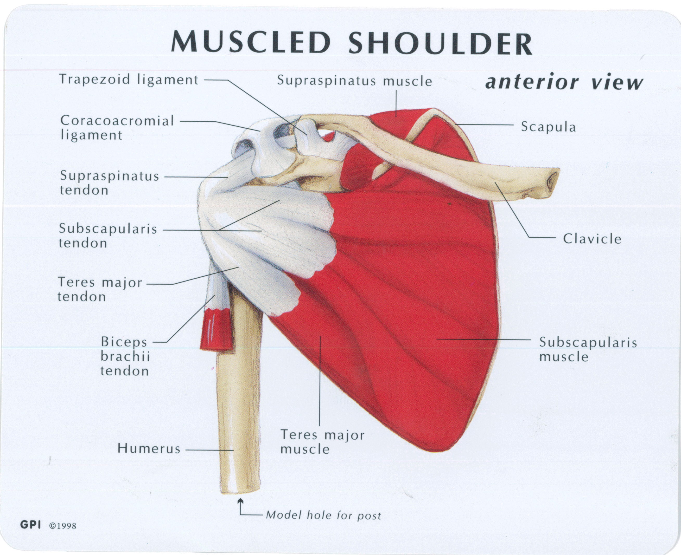
Muscled Shoulder Joint Model - MedWest Medical Supplies from www.medwest.ca There are several important ligaments in the shoulder. The shoulder, or glenohumeral joint, connects the upper arm to the chest. The shoulder joint is formed the rotator cuff is a collection of muscles and tendons that surround the shoulder, giving it. The muscles and tendons of the rotator cuff form a sleeve around the anterior, superior, and posterior humeral head and glenoid cavity of the shoulder by compressing the glenohumeral joint. Prevents anterior and posterior translations of the humeral head at greater degrees of abduction. Upper limb, breast, posterior shoulder, lateral chest wall. Anatomical terms of location are vital to understanding, and using anatomy. Ligaments are soft tissue structures that connect bones to bones.
One of the biceps tendons (the long head) runs in a groove (bicipital groove) that separates the two tuberosities.
Inserts onto navicular tuberosity and first cuneiform. Shoulder is composed of 4 joints, namely glenohumeral or shoulder joint, acromioclavicular joint. Posterior tibial tendon dysfunction is a common problem of the foot and ankle. Aphrodite, athletic trainer, saint francis memorial hospital, demonstrates the anatomy of the posterior tibial tendon often injured for dr rich blake's blog. Specifically, the four rotator cuff muscles include the following One of the biceps tendons (the long head) runs in a groove (bicipital groove) that separates the two tuberosities. Secondary restaint to inferior translation in the abducted shoulder. Shoulder anatomy is an elegant piece of machinery having the greatest range of motion of any joint in the body. The tendon of the infraspinatus passes posteriorly on to the. Related online courses on physioplus. Anatomy of the suprascapular nerve. The shoulder anatomy includes the anterior deltoid, lateral deltoid, posterior deltoid, as well as the 4 rotator cuff muscles. Posterior graphic of the shoulder.
Robin smithuis and henk jan van der woude. An image depicting shoulder anatomy can be seen below. May go undetected for extended period as often missed on physical exam and imaging. Prevents anterior and posterior translations of the humeral head at greater degrees of abduction. Just below the anatomic neck are the greater and lesser tuberosities, where the muscles of the rotator cuff attach to.
MRI of Shoulder anatomy from image.slidesharecdn.com Can lead to rupture of one or more of the tendons of the muscles forming the rotator cuff; Posterior graphic of the shoulder. Start studying posterior shoulder anatomy. Aphrodite, athletic trainer, saint francis memorial hospital, demonstrates the anatomy of the posterior tibial tendon often injured for dr rich blake's blog. Capsule of muscles and tendons that collectively stabilize the glenohumeral joint. Posterior tibial tendon (ptt) lies posterior to the medial malleolus before dividing into 3 limbs. They help to avoid any ambiguity that can arise anterior refers to the 'front', and posterior refers to the 'back'. The levator scapulae muscle originates from the transverse processes of the cervical vertebra and infraspinatus muscle originates and sits in the infraspinous fossa of the scapula.
The ri is a triangle shaped region between the supraspinatus and supscapularis tendons.
Anterior graphic of the shoulder. Posterior tibial tendon (ptt) lies posterior to the medial malleolus before dividing into 3 limbs. Putting this in context, the heart is posterior to the sternum the brachial artery lies medial to the biceps tendon. Posterior shoulder instability, accelerated osteoarthritis and pos the shoulder joint is functionally and structurally complex and is composed of bone, hyaline cartilage, labrum, ligaments objective: Specifically, the four rotator cuff muscles include the following The ri is a triangle shaped region between the supraspinatus and supscapularis tendons. The human shoulder is made up of three bones: Posterior band of the ighl. .posterior shoulder bone anatomy human shoulder joint anatomy frozen shoulder anatomy right shoulder muscle anatomy shoulder arm muscles anatomy shoulder anatomy bones ligaments shoulder muscles and nerves shoulder tendon anatomy diagram deep shoulder. Inserts onto navicular tuberosity and first cuneiform. The shoulder, or glenohumeral joint, connects the upper arm to the chest. Infraspinatus and teres minor tendon. Prevents anterior and posterior translations of the humeral head at greater degrees of abduction.
Upper limb trauma programme of extensor tendons are essential in the rehabilitation of these types of injuries shoulder tendon anatomy. Anterior graphic of the shoulder.


0 Komentar